
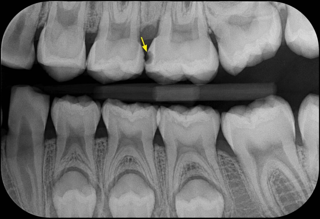
Your pediatric dental team just recommended bitewing X-rays for your child, and as a parent, you are naturally wondering if they are truly necessary. It is a common question among Hawaii parents who want to balance medical safety with comprehensive, protective oral care. Here is the clinical reality: visual dental exams alone detect only a limited fraction of cavities that form between teeth. Without these targeted, high-resolution diagnostic images, the vast majority of childhood tooth decay goes completely unnoticed until it is large enough to cause visible structural damage, deep enamel destruction, or throbbing pain.
By the time a cavity becomes detectable to the naked eye during a standard mirror examination, the treatment required is often far more invasive, potentially involving extensive composite restorations, pediatric crowns, or nerve therapies. This comprehensive guide explains exactly what bitewing X-rays reveal, how often developing children actually need them, and why modern digital dental technology makes them safer and faster than ever before. At Pedodontic Associates, we have been partnering with Hawaii families since 1977 to provide gentle guidance and clear education regarding every aspect of your child’s dental imaging.
What Bitewing X-Rays Show That Your Dentist Can’t See
A bitewing X-ray captures a highly detailed snapshot of the back teeth (pre-molars and molars). It focuses specifically on the crowns of the teeth—the visible portion that rises above the gumline—and the underlying supporting bone levels surrounding them. During the diagnostic process, your child bites down gently on a small wing-shaped tab that aligns the digital sensor flat against the interior cheek, which is exactly how these specialized images earned their name.
While the image captures teeth from both the upper and lower jaws simultaneously to evaluate how they meet, its primary diagnostic purpose is to help our team identify and diagnose cavities forming between adjacent teeth within the exact same dental arch.
Targeting Banned Hiding Spots: “Interproximal” Decay
This technology is essential because the majority of hidden childhood cavities start in the “interproximal” spaces—the tight, compressed areas where adjacent back teeth physically press against one another. In Hawaii’s warm, tropical climate, families regularly enjoy local treats like shave ice, sticky fruits, and sweet juices. When these sugars accumulate in tight dental spaces, they provide the ideal fuel for acid-producing bacteria to thrive. Because toothbrush bristles cannot physically slide into these compressed spaces, plaque accumulation goes completely undisturbed.
By the time a parent or dental provider can see a cavity between the molars without an X-ray, the decay has typically bypassed the thin, protective outer enamel and burrowed deep into the softer dentin layer underneath. Catching this breakdown early via a routine digital bitewing can mean the difference between a simple, non-invasive treatment or a small filling and an expensive, complex pediatric crown.
Comparing Common Pediatric X-Rays
| X-Ray Type | Primary Focus Area | Best Clinical Use Case for Keiki |
|---|---|---|
| Bitewing | Between the upper and lower back teeth (pre-molars and molars). | Detecting “hidden” early-stage cavities and checking existing filling margins. |
| Panoramic | Full jaw, sinuses, jaw joints, and all upper/lower arches in one wide view. | Orthodontic development planning, tracking impacted teeth, and wisdom teeth alignment. |
| Periapical | The entire tooth structure from the visible crown down to the root tip. | Evaluating deep localized trauma, checking for abscesses, and tracking root infections. |
How Often Your Child Actually Needs These X-Rays
The American Academy of Pediatric Dentistry (AAPD) establishes clear consensus guidelines for dental imaging frequency based on a child’s specific susceptibility to decay. For children presenting with active clinical decay or identified as carrying a high risk for cavities, the AAPD recommends a posterior bitewing examination at 6 to 12-month intervals. Conversely, for low-risk pediatric patients with no active clinical caries and optimal home habits, that interval safely extends to every 12 to 24 months.
This professional timeline is dictated by one primary biological factor: the rapid speed at which pediatric tooth decay travels. Primary baby teeth possess a significantly thinner outer layer of enamel compared to permanent adult teeth. A tiny spot of demineralization that might take a few years to progress into a serious structural issue in an adult can rapidly burn through a child’s thin enamel, entering the pulp and becoming a painful dental emergency in less than a year.
Evaluating the Individual Smile Milestone
At Pedodontic Associates, we do not implement a rigid, “one-size-fits-all” operational rule based strictly on age. Instead, we customize our diagnostic approach to match your child’s unique oral anatomy. We carefully assess when a child’s back primary molars physically shift and lock tightly flush against one another, creating contact points that block direct visual inspection.
While this structural closing of spaces frequently occurs around age four, our highly trained clinical staff often captures bitewing radiographs on very capable older 3-year-olds if their posterior teeth are already touching adjacently. If the spaces remain wide open and completely visible to a manual mirror exam, we safely defer the radiographs to a later visit.
The Real Numbers on Radiation Safety: Facts vs. Fear
It is entirely natural for parents to feel protective when it comes to radiation exposure. However, the radiation levels associated with modern pediatric digital technology are incredibly low, making the risk of leaving hidden decay undetected far greater than the scan itself. A standard digital bitewing series exposes your child to a negligible fraction of radiation—approximately 0.005 millisieverts (mSv).
Putting Exposure into a Hawaii Context:
- Natural Background Radiation: Simply living in our beautiful islands for two days exposes your child to more natural background radiation from the tropical sun and local volcanic soil than a complete digital dental X-ray series.
- Pacific Air Travel: A single round-trip commercial flight from Honolulu to Los Angeles exposes your child’s body to roughly 0.035 mSv of cosmic radiation—which is about seven times more exposure than a standard set of bitewing dental pictures.
- Everyday Dietary Sources: Due to naturally occurring potassium-40 found in fresh produce, eating approximately 50 standard bananas provides the exact same baseline radiation exposure as a modern digital dental radiograph.
Our practice strictly enforces the “ALARA” principle (As Low As Reasonably Achievable) across our Aiea, Honolulu, and Maui locations. We utilize specialized, high-speed digital sensors that instantly record images while requiring up to 80% less radiation than old-fashioned dental film. Furthermore, we protect every child using lead-lined safety aprons equipped with built-in thyroid collars, combined with precise rectangular collimation to completely shield developing tissues from stray exposure.
What Happens During Your Child’s Appointment
The actual digital X-ray capture process is remarkably efficient, typically taking only 3 to 5 minutes to complete from start to finish. To ensure maximum efficiency and child comfort, our highly trained clinical staff and dental assistants complete the imaging capture process. Our team of skilled pediatric dentists—which includes several pediatric specialists—then meticulously evaluate the completed radiographs to form a precise diagnostic map.
During the session, a compassionate assistant will place a small, cushioned digital sensor inside your child’s cheek. Your child will be asked to bite down gently on the attached “wing” tab to hold the sensor perfectly steady. The compact dental imaging head, placed on the outside of their cheek, clicks an image in less than half a second.
Using “Tell-Show-Do” behavioral Guidance
We understand that a small percentage of young children experience a sensitive gag reflex or feel anxious about placing dental sensors inside their mouths. Because our team consists exclusively of specialized pediatric dentists who completed years of post-doctoral residency training, we utilize the evidence-based Tell-Show-Do method to make the process completely stress-free:
- Tell: We introduce our low-radiation dental X-rays using playful, kid-friendly descriptions. We explain that our “magic tooth camera” needs to take a quick, special picture to see how strong their superhero teeth are growing inside their gums.
- Show: We let the child physically touch the plastic sensor flap with their finger and see the imaging machine up close so they understand it contains no sharp edges and isn’t scary.
- Do: Once the child is laughing and comfortable, our team works with swift precision to capture the clean digital picture on the very first try, minimizing the time the sensor rests in the mouth.
What Your Child’s X-Rays Actually Reveal
Digital bitewing X-rays provide our clinical team with a highly accurate structural map of your child’s developing mouth, extending far beyond basic cavity detection:
- Evaluation of Existing Restorations: They show our doctors if older pediatric crowns or standard dental fillings are beginning to experience microscopic breakdown at the margins, preventing secondary decay underneath.
- Tracking Tooth Eruption Pathways: They allow us to accurately monitor how permanent adult teeth are moving beneath the surface and verify if there is sufficient space within the growing jaw arch to accommodate them.
- Monitoring “Watch” Areas: Bitewings allow us to map out “incipient” lesions—tiny, early-stage areas of enamel demineralization that haven’t broken down into physical holes yet. By spotting these spots early, we can often intervene with conservative, non-invasive enamel-reversing therapies like Curodont™ to avoid traditional drilling completely.
Parent Reviews: Trusting the Experts
Don’t just take our word for it—Hawaii parents trust our team to provide safe, effective diagnostic care:
Read Jon D.‘s review of Pedodontic Associates – Kahala on YelpRead Di S.‘s review of Pedodontic Associates – Kahala on Yelp
Proactive Care for a Lifetime of Smiles
Waiting until a child actively complains of a physical toothache before taking dental radiographs is a risky approach. By the time a hidden interproximal cavity causes constant pain, the decay has typically penetrated deep into the inner nerve chamber of the tooth. This often transforms a simple preventive fix into a complex, emergency situation requiring a nerve treatment or a total tooth extraction.
Routine, low-radiation digital bitewing X-rays allow our team to identify structural modifications when they are small, manageable, and highly treatable. To make this essential diagnostic care fully accessible for busy local families, our offices provide convenient Saturday appointment options, ensuring your child never has to miss school to maintain a healthy smile.
Protecting Accessible Care Options
We are firmly committed to providing the highest tier of pediatric care to all keiki across our islands. Our administrative team works directly with major local insurance networks to maximize your benefits and keep out-of-pocket costs transparent. We proudly accept and process claims with Hawaii Dental Service (HDS)—the state’s largest standalone dental provider—alongside HMSA, HMAA, and Hawaii Quest (Medicaid) community dental programs.
Are you ready to establish a proactive dental home and evaluate your child’s developing smile? Contact Us today to schedule a comprehensive pediatric exam at our Aiea, Honolulu, or Maui office!
Medical & Legal Disclaimer: The content on this blog is provided for general informational purposes only and is not intended as medical advice, diagnosis, or treatment. Always seek the advice of a qualified pediatric dentist regarding your child’s specific oral health needs. If your child is experiencing a medical emergency, including severe facial swelling, call 911 or proceed to the nearest emergency room immediately.